Expand Access to Physician-Reviewed Retinal Screening
Orvos Healthcare is dedicated to advancing eye care through retina screening with innovative technology and strategic partnerships. Our collaboration with leading non-mydriatic fundus camera providers enables us to deliver comprehensive remote retina screening services across the United States. By leveraging cutting-edge imaging technology, we provide healthcare professionals with the tools necessary for accurate and timely diagnosis of retinal conditions. Our focus of quick turnaround times ensures that patients receive the care they need without delay, significantly enhancing clinical workflows. Additionally, our services play a crucial role in improving care metrics, ultimately contributing to better health outcomes for our patients. Together, we are transforming the landscape of eye care and making a meaningful impact on public health.
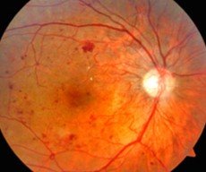
AI vs AI with Human in the loop
Human-in-the-loop screening combines AI efficiency with physician oversight. The AI highlights possible concerns, while a qualified physician reviews the images, confirms or modifies the findings, evaluates additional visible abnormalities, and recommends the appropriate level of follow-up.
Conditions Diagnosed
Leveraging Human In The Loop
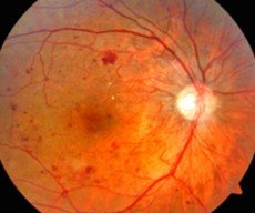
Diabetic Retinopathy - DR
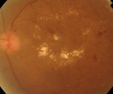
Diabetic Macular Edema - DME
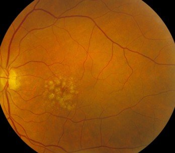
Macular Degeneration - AMD
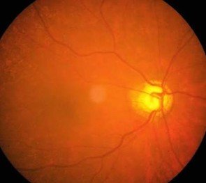
Glaucoma
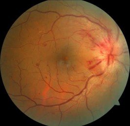
Hypertensive Retinopathy
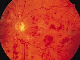
Retinal Vein Occlusion
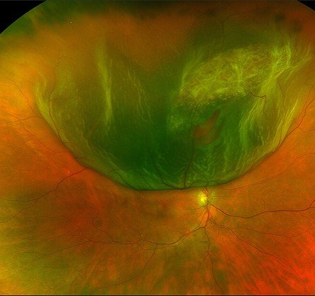
Retinal Tear or Detachment
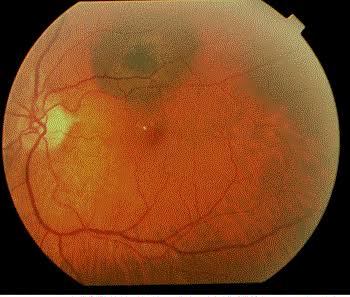
Suspicious Retinal Lesion
How the Process Works
Who We Serve
Flexible retinal screening and physician-review services designed for diverse care settings.